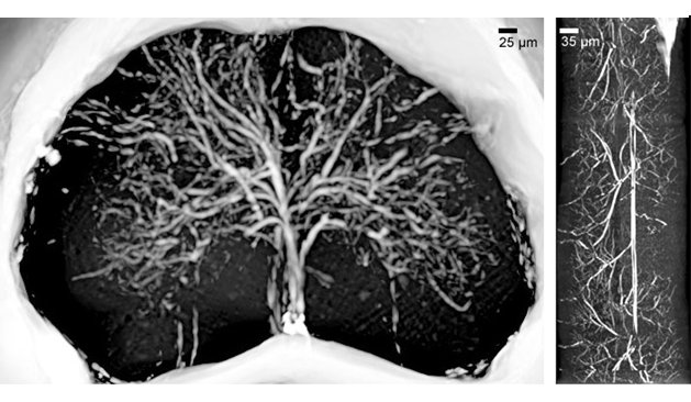

Figure 1: (a) X-ray phase contrast microtomography of the lumbar-sacral region of the spinal cord (size: 1.2 mm). (b) Reconstructed volume of the lumbar-sacral region aft er administration of the MICROFIL contrast agent. The image pixel size is 3.5 μm. (c) Longitudinal view of (b), 1 mm long.
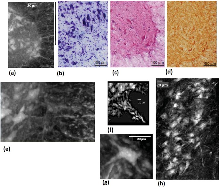
Figure 2: Neural population: (a) White/grey matter interface of a thick slab selected in the anterior horn of the lumbar-sacral spinal cord. (b,c,d) For comparison, microscope histological images: (b) Histology (Nissl staining), (c) Histology (He/eo staining) and (d) Immunohistochemical analysis of SMI-32, a marker of motor neurons. (e) zoom of the with/grey matter anterior horn interface. (f) Magnification of a single neuronal cell. (g) Image of one nerve fibre at the interface with the grey matter. (h) Longitudinal view (length: 0.5 mm) of the sample at the same interface.
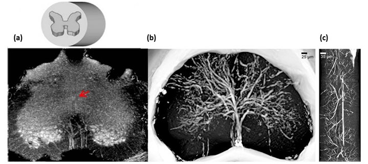
(a) X-ray phase contrast microtomography of the lumbar-sacral region of the spinal cord (size: 1.2 mm). (b) Reconstructed volume of the lumbar-sacral region aft er administration of the MICROFIL contrast agent. The image pixel size is 3.5 μm. (c) Longitudinal view of (b), 1 mm long.